INNOVATIVE SURGICAL TECHNIQUES / CHILDREN
PEDIATRIC SPINAL TUMORS

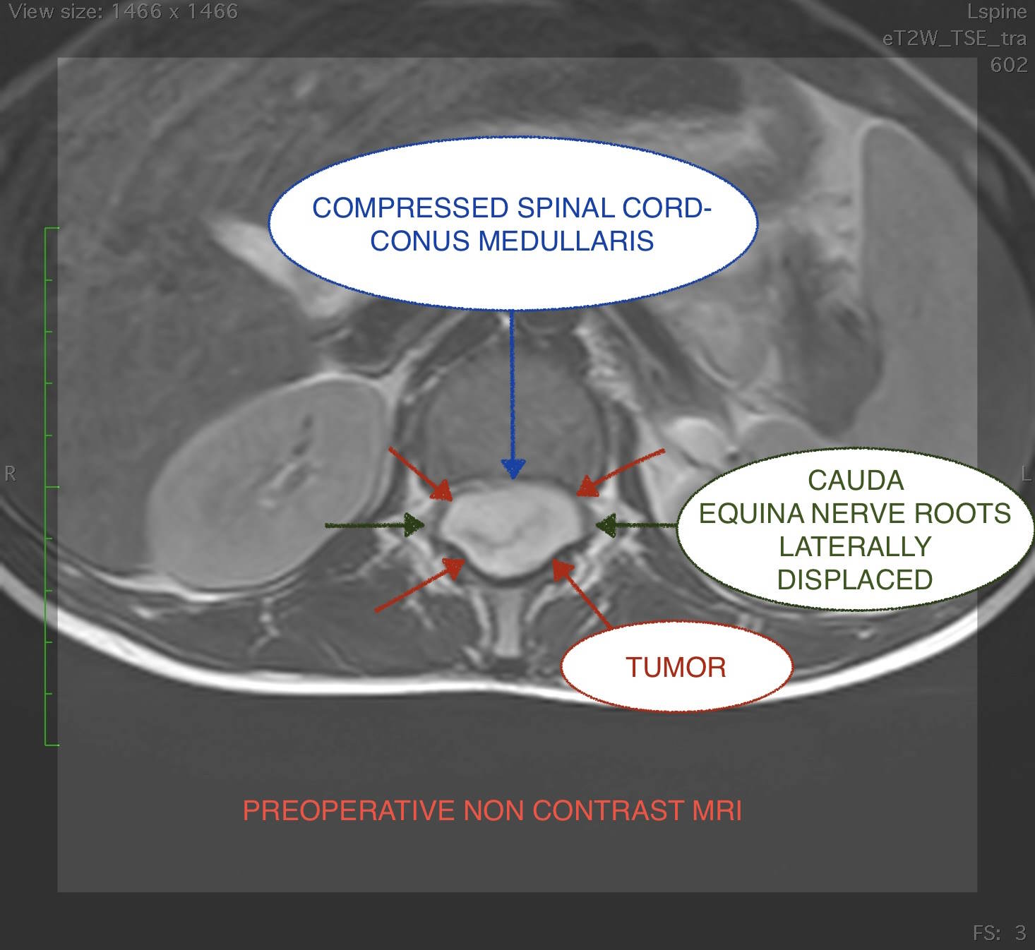
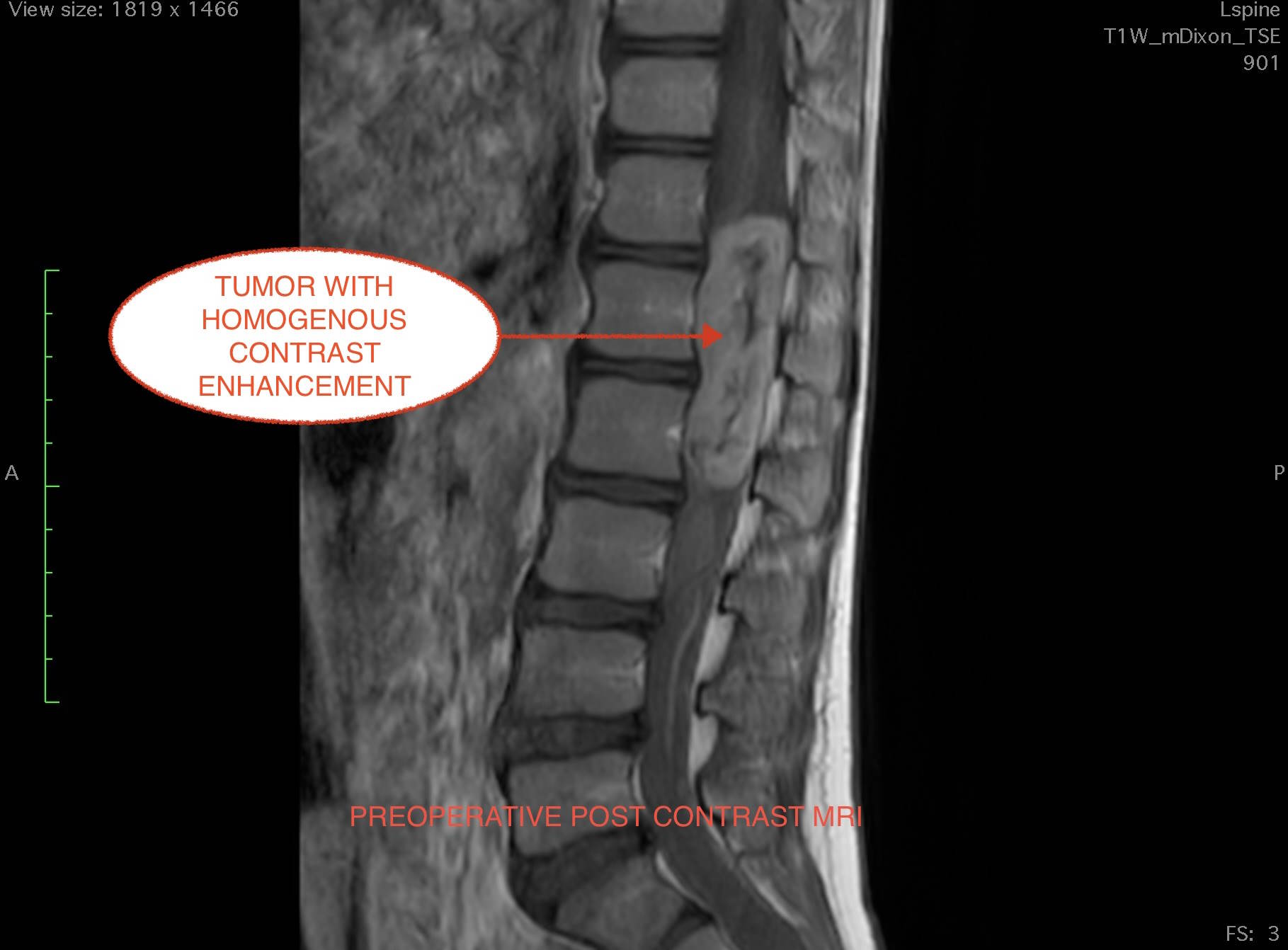
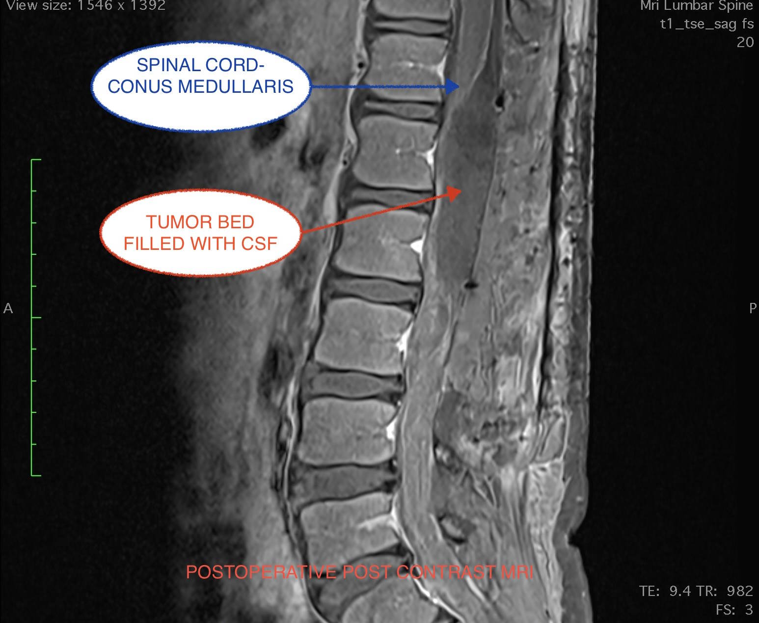
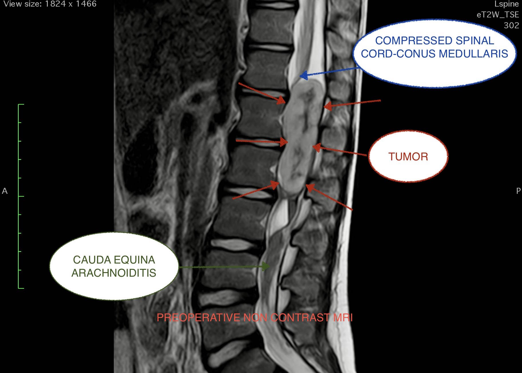
We present the case of a 12-year-old boy, presenting with symptomatology of persistent lower thoracic back pain and lower extremities tingling. He underwent lumbar spine MRI which revealed the presence of a large intradural extramedullary tumor, with homogenous contrast uptake extending to levels T12 to L2. The tumor was significantly compressing the conus medullaris and was laterally displacing the cauda equina nerve roots, which also demonstrated arachnoiditis-like appearance caudally to the tumor. The patient underwent also whole neuraxis MRIs with and without contrast with no other pathology revealed.

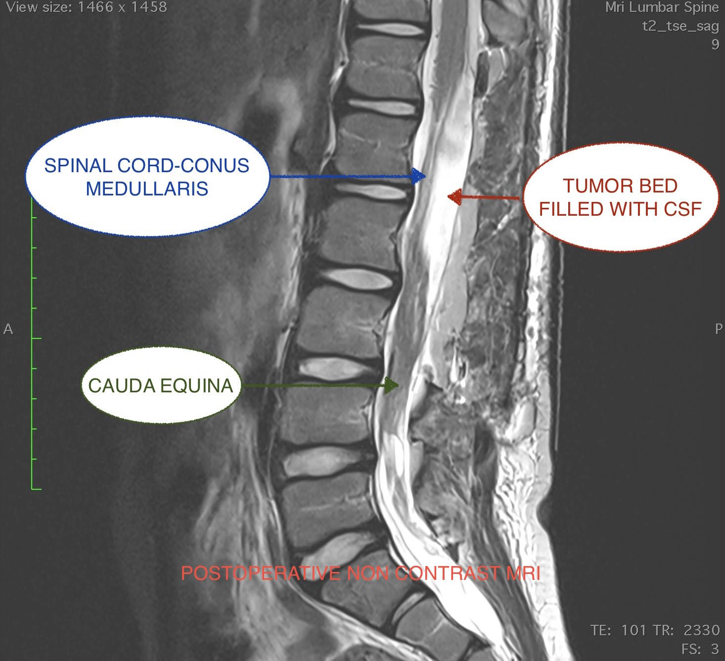
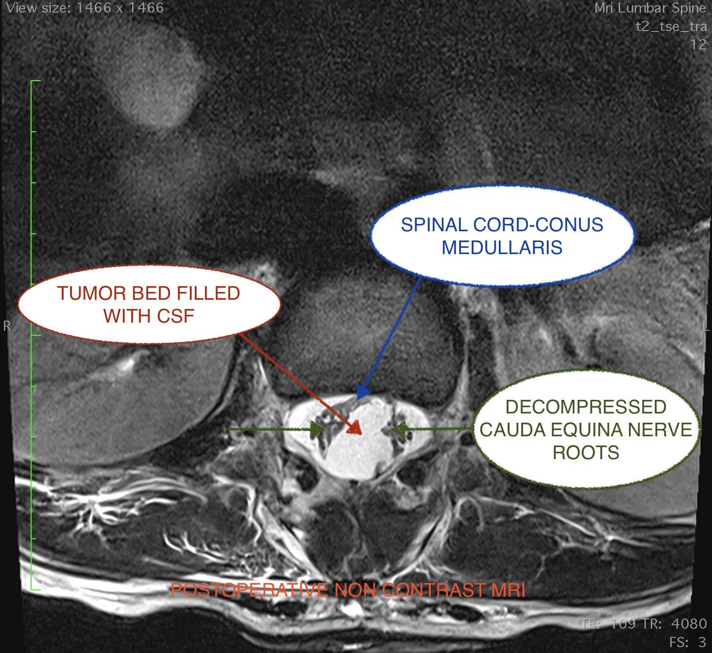
Treatment included total surgical removal and tumor biopsy. The procedure was performed under continuous intraoperative neuromonitoring of the conus medullaris and cauda equina nerve roots. The patient was placed prone onto the operating table and after T12/L1/L2/L3 laminectomies, the dura was incised and the tumor visualized. Cerebrospinal fluid (CSF) sample was obtained and sent to cytology lab. The tumor was found to be firmly attached to the filum terminale, which was also infiltrated by the tumor. After neuromonitoring confirmation, the filum terminale was cauterized and incised, a maneuver which facilitated the total excision of the tumor. Watertight dura closure was performed, and the wound was closed in layers. Electrophysiological recordings were stable throughout the procedure. The patient emerged from anesthesia into the operating room and was extubated without any complication. He was then transferred to neurosurgical ward. He was able to walk and urinate in the first postoperative day and he was discharged home on the third postoperative day, with no neurologic deficit. On the second postoperative day he underwent lumbar spine MRI pre and post contrast, with no residual tumor demonstrated.

Biopsy revealed myxopapillary ependymoma WHO Grade 2. CSF cytologic examination was negative for malignant cells. After oncologist consultation, the young patient will need no supplementary treatment, and he will undergo scheduled follow-up spine MRIs for the following years.

Skull base tumor with concomitant fistula in a 3-year-old patient

We treated the rare case of a young boy who presented with a small skin lesion on the forehead between the orbits. This skin lesion had never been assessed until a few months ago when our young patient began suffering episodes of fluid effusion from the area. He underwent a brain MRI that revealed a benign skull base tumor (dermoid cyst) that had caused the bone to become thinner and had created a fistula to the patient’s nose. This is a very serious condition because it disrupts the isolation of the brain, from the external environment possibly resulting in a severe infection of the nervous system that may threatent the patient neurological integrity and even his/her life.

We performed a cunolox surgery that includes a craniotomy in the frontal and orbital area, and then completely removed the tumor as well as the fistula.

The young patient was discharged after only a few days of hospitalization in excellent neurological condition.

Dermoid cyst with intradural extension and tract

Radical excision of the cyst along with its adherence to the cerebral falx

Spinal cord tethering with concomitant meningocele and lipoma in a 6 month-old infant

Spinal cord tethering accompanied by meningocele is usually recognized immediately after birth and requires surgical treatment in the first months of one’s life in order to protect leg functionality, as well as bladder and bowel control. We treated a 6-month-old infant who further presented with a lipoma (a benign tumor) inside the spine, in contact with the spinal cord and nerve roots, significantly increasing the surgical difficulty. The increased difficulty is due to the anatomical relationship of the tethered spinal cord with the lipoma.

Our young patient was underwent to microsurgical (using surgical microscope), removal of the lipoma and detethering of spinal cord  with aesthetic restoration.

We are confident that she will have a normal life without any neurological deficits.

Post traumatic C1 and C2 fracture with C1/C2 subluxation

We treated and extremely urgent case of a 5 years old girl, who was transferred to the Emergency Department of our Hospital after a fall from standing height which resulted in cervical spine injury. On clinical examination, patient was neurologically intact with severe cervical spine tenderness and right lateral flexion/right rotation torticollis.

She underwent cervical spine CT scan which revealed oblique odontoid tip fracture, anterior arch of C1 fracture, anterolisthesis of C2 over C3 and rotational subluxation of 25 degrees of C1 over C2.

Treatment included cervical immobilization with hard collar and urgent surgical operation with open reduction of C1/C2 subluxation followed by posterior instrumented fusion C1/C2/C3 with lateral mass screws and 2 rods under continuous intraoperative neuromonitoring